

Hyperspectral detection technology
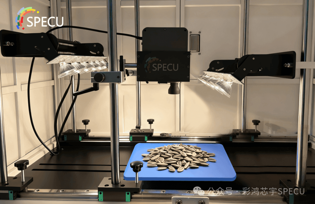
SPECU hyperspectral camera module for sunflower seed inspection
In precision detection, different technologies support the development of multiple industries through their distinct strengths. Hyperspectral technology stands out for its dual-dimensional analysis capability combining imaging and spectroscopy. This article compares it with detection techniques such as Raman spectroscopy and fluorescence spectroscopy, outlining their respective application scenarios and core differences.
Hyperspectral technology combines imaging and spectral analysis to capture both spatial images and continuous spectral curves of a target at the same time. Its core advantage is “non-destructive and visual + precise composition analysis” — it can both observe a target's morphology and use spectral features to infer material composition and content, such as measuring sugar content in fruits and vegetables in agriculture or identifying water pollutants in environmental protection.
1
Hyperspectral vs. Raman spectroscopy detection techniques
Raman spectroscopy uses the “inelastic scattering of light” (the Raman effect) to analyze the vibrational energy levels of molecules. Its core is “molecular structure fingerprinting”: it requires no sample pretreatment and can measure gases, liquids, and solids. Hyperspectral methods, by contrast, need to be combined with spatial imaging and focus on “composition distribution analysis across areal regions.”
In application, Raman spectroscopy is well suited to trace-component analysis in the lab (e.g., drug purity identification), whereas hyperspectral imaging is better suited to large-area on-site inspection (e.g., crop monitoring in farmland and simultaneous screening of surface defects and composition on industrial products).
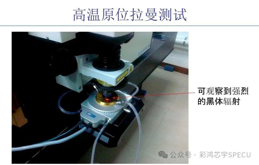
Image source: Sohu account – Guoxin Zhongye Institute of Science and Technology
2
Hyperspectral vs. ground-object spectral detection technology
Surface-object spectroscopy focuses specifically on surface objects (such as vegetation, soil, and rock) and measures their reflectance/emission spectra within specific wavelength ranges. Its core function is “spectral feature acquisition at a single point or over an area,” with no imaging capability. Hyperspectral technology, by contrast, combines “surface-object spectroscopy + spatial imaging” and can show differences in the spatial distribution of spectra.
Ground feature spectra are mainly used for remote sensing data calibration (such as ground-truth measurements for satellite remote sensing) and for building spectral libraries of geological samples. Hyperspectral data can directly generate spectral imagery maps, making it suitable for precision agriculture (distinguishing crop nutrient differences across plots) and ecological monitoring.
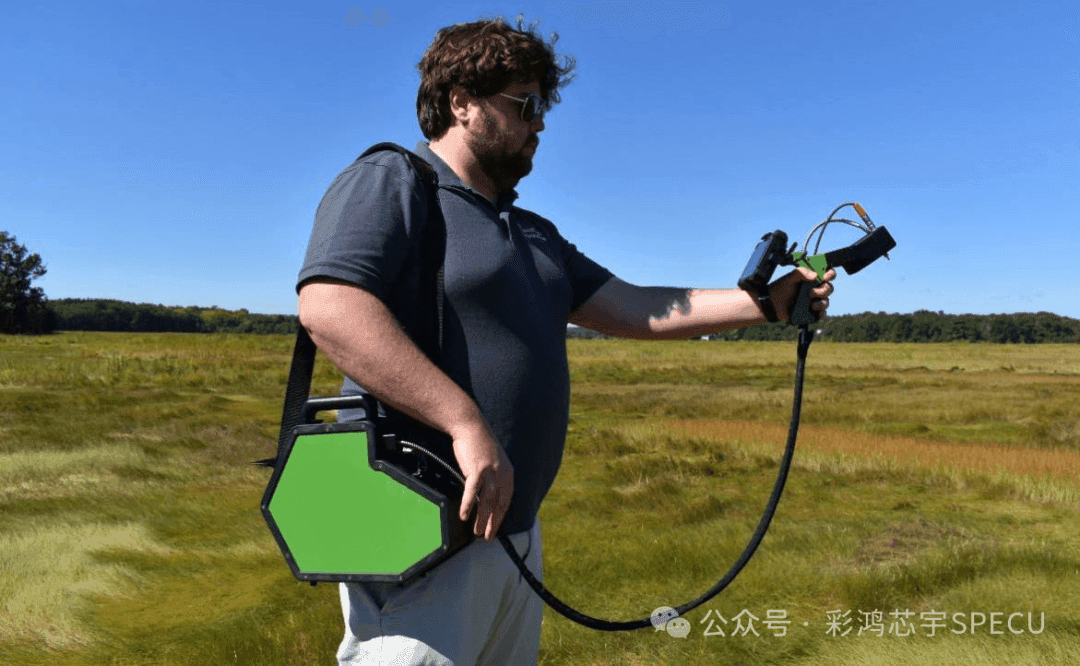
Image source: Baidu
3
Hyperspectral vs. fluorescence spectroscopy detection technology
Fluorescence spectroscopy uses excitation light to induce fluorescence in a substance and analyzes the relationship between fluorescence intensity and wavelength. Its core purpose is "high-sensitivity detection of fluorescent substances," and it depends on the substance's fluorescence properties. Hyperspectral techniques require no excitation and directly capture a substance's own reflection/emission spectra, giving them broader applicability.
Fluorescence spectroscopy is mainly used for detecting trace fluorescent substances (such as screening for fluorescent whitening agents in food and detecting algal toxins in water); hyperspectral imaging is better suited to non-fluorescent scenarios (such as internal quality inspection of fruits and vegetables).
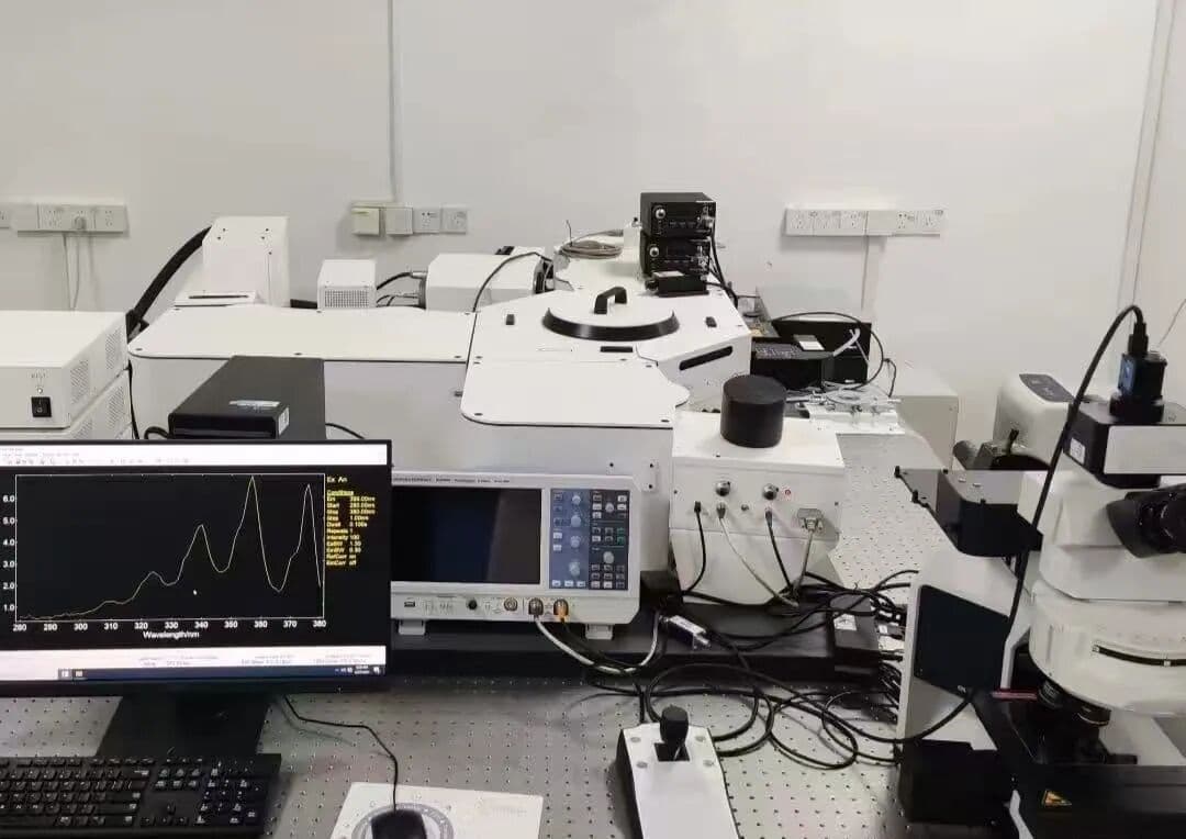
Image source: Xiaohongshu
4
Hyperspectral vs. fiber-optic spectral detection technology
Fiber-optic spectroscopy uses optical fiber as the transmission medium to collect spectral information from a target. Its core capability is “long-distance, microscale spectral acquisition” (for example, detecting substances inside industrial pipelines or analyzing microscale regions of living biological tissue), but it can only obtain “point/line” spectra and has no imaging capability. Hyperspectral can achieve “area imaging + spectral analysis.”
Fiber-optic spectroscopy is suited to single-point detection in confined spaces and hazardous environments (for example, analyzing corrosion products inside oil and gas pipelines); hyperspectral imaging is suited to visualizing compositional distribution over large areas (for example, inspecting compositional uniformity on industrial wafer surfaces).
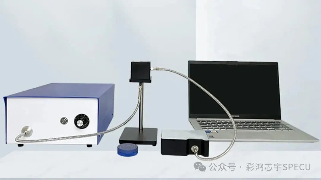
Image source: Zhihu
5
Hyperspectral vs. nuclear magnetic resonance detection technology
Nuclear magnetic resonance (NMR) uses the resonance of atomic nuclei in a magnetic field to analyze the molecular structure and composition of materials. Its core strength is “non-destructive, high-resolution molecular structure analysis,” but the equipment is bulky and expensive, and samples must be placed in a specific magnetic field environment. Hyperspectral equipment is more portable and enables on-site testing without complex environmental control.
NMR is mainly used for precise molecular structure studies in laboratories (such as drug molecule design and molecular-level analysis of food components); hyperspectral imaging is suitable for rapid on-site testing (such as quality grading at agricultural harvest sites and in situ screening of environmental pollutants).
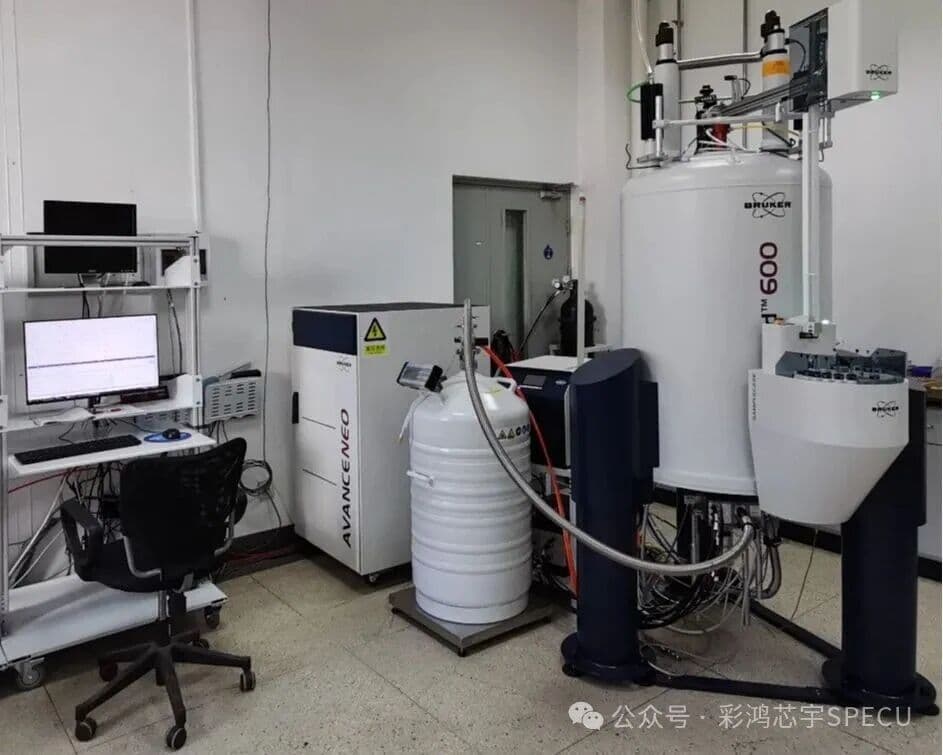
Image source: School of Physics and Materials Science, Changji University
6
Hyperspectral vs. broad-spectrum detection technology
Broad-spectrum coverage spans a wide wavelength range (typically wider than hyperspectral), but spectral resolution is low (large band spacing, no continuous spectral curve). Its core feature is “wide wavelength coverage” (e.g., full-band scanning across ultraviolet, visible, and near-infrared); hyperspectral systems have high spectral resolution and can capture subtle spectral differences.
Broadband spectroscopy suits applications with low spectral resolution requirements, such as water turbidity testing and monitoring total atmospheric pollutant concentrations; hyperspectral spectroscopy suits applications that require precise discrimination of material composition, such as distinguishing different types of plastic and spectral diagnosis of early-stage cancer tissue.
7
Hyperspectral vs. ultrasonic testing technology
Ultrasonic testing uses differences in how ultrasound propagates through different media to detect internal defects and structures in materials. Its core capability is “deep, nondestructive probing of internal structures” (for example, detecting defects in metal welds and ultrasound imaging of human organs); hyperspectral imaging cannot penetrate materials and can analyze only surface and shallow-layer composition.
Ultrasound is mainly used for internal structure inspection (such as internal crack detection in industrial castings and prenatal fetal ultrasound examinations); hyperspectral imaging focuses on surface and shallow-layer composition analysis (such as spectral diagnosis of skin lesions and mold detection on food surfaces).
Image source: Baidu Baike
8
Hyperspectral vs. near-infrared spectroscopy detection technology
Near-infrared spectroscopy focuses on the near-infrared band (780–2500 nm) and uses the absorption characteristics of substances in this band to analyze composition. Its core advantage is fast, low-cost detection, but it covers only a single band and provides limited spectral information; hyperspectral technology can cover multiple bands, including ultraviolet, visible, and near-infrared, and provides richer spectral information.
Near-infrared spectroscopy is suitable for rapid, large-batch screening (such as bulk testing of grain moisture content and preliminary screening of ingredient uniformity in pharmaceutical tablets); hyperspectral analysis is suited to scenarios that require detailed multi-band analysis (such as simultaneous detection of multiple pesticide residues in agricultural products and identification of multiple paint layers on cultural relics).
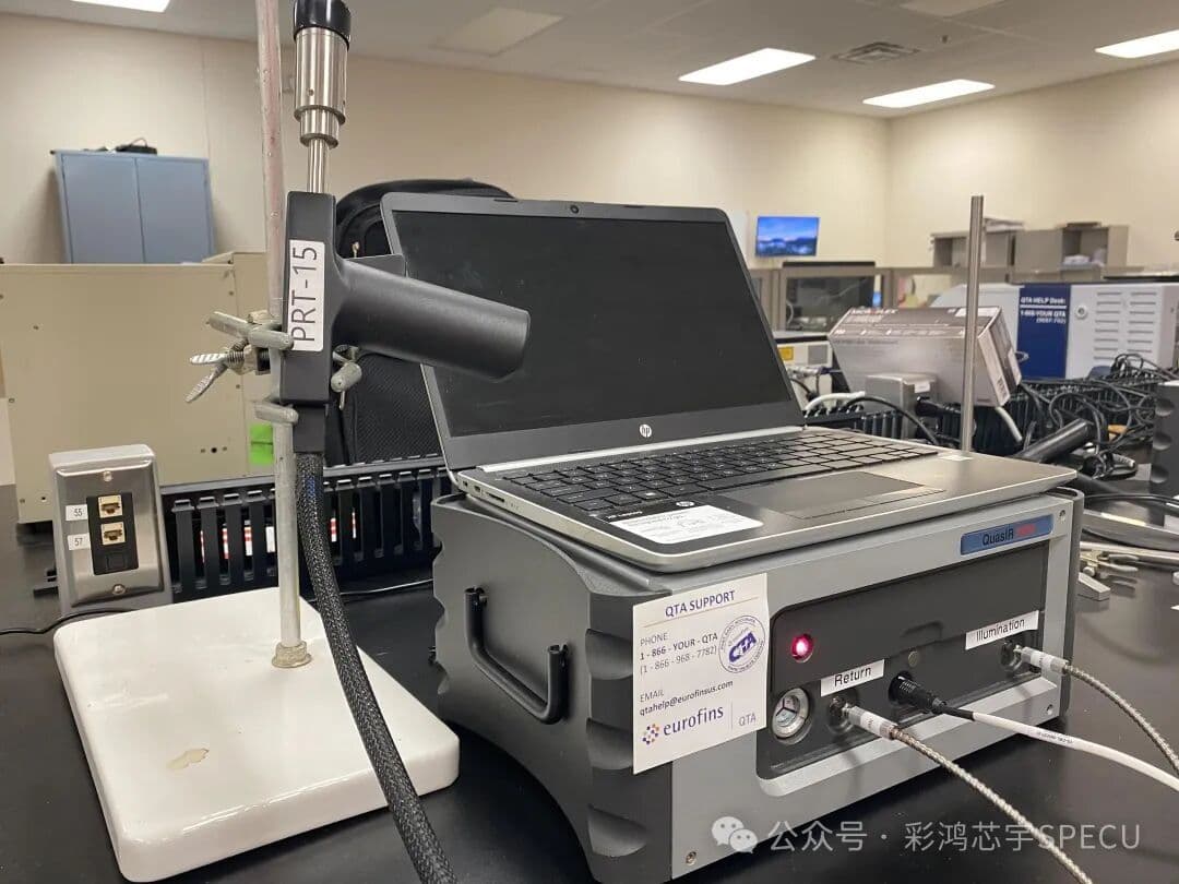
Image source: Zhihu
9
Hyperspectral vs. microwave detection technology
Microwave inspection uses the penetration and reflection properties of microwaves (wavelength 1 mm–1 m) to inspect internal structure and moisture content. Its core strengths are “penetrating non-metallic materials and enabling rapid inspection” (for example, moisture monitoring in grain storage and internal defect detection in concrete); hyperspectral imaging has no penetration capability and does not rely on microwave properties.
Microwave technology is suitable for internal inspection of non-metallic materials (such as detecting internal voids in wood and foreign objects in food packaging); hyperspectral technology is suitable for precise analysis of surface composition (such as detecting coating composition on industrial product surfaces and mapping heavy metal content in environmental soil).
10
Hyperspectral vs. terahertz detection technology
Terahertz inspection uses the properties of terahertz waves (wavelength 0.3–3 mm, between infrared and microwave) for imaging. Its core advantages are non-destructive penetration and low energy: it can penetrate nonmetallic materials such as paper, plastic, and fabric, and is non-ionizing to biological tissue. Hyperspectral imaging has no penetration capability and focuses on spectral composition analysis of surface and shallow materials.
In terms of applications, terahertz inspection is mostly used for security screening (such as identifying concealed dangerous goods in packages), semiconductor quality inspection (detecting internal chip defects), and pharmaceutical packaging integrity testing; hyperspectral imaging, by contrast, is better suited to scenarios that require precise analysis of material composition, such as testing the internal quality of agricultural products (sugar content and acidity) and identifying the composition of environmental pollutants.
Image source: Baidu Baike
Summary
Laboratory precision testingNMR analyzes molecular structures, and high-performance liquid chromatography (HPLC) accurately quantifies components. Both require a laboratory environment, have long testing times, and are suitable for research and precision analysis.
Single-point rapid testing: Raman spectroscopy for rapid material identification, near-infrared spectroscopy (NIRS) for batch moisture measurement, and fiber-optic spectroscopy for micro-area composition analysis are all “point/line” measurements, so they cannot show composition distribution over large areas.
Special-function categoryFluorescence spectroscopy specifically detects fluorescent substances (such as fluorescent whitening agents in food); broad-spectrum scanning quickly measures pollutant concentrations; polarization imaging distinguishes camouflage or measures roughness. It is efficient but has narrow applicability.
Penetration detection category:Ultrasonic testing detects internal defects (such as cracks in metal), while microwaves pass through nonmetallic materials to inspect the interior (such as voids in wood). The key is “seeing inside”; these methods cannot analyze material composition.
Ground object-specific category:Ground object spectroscopy collects vegetation and soil spectral data to provide “ground truth” for remote sensing calibration. It has no imaging capability and cannot show spatial distribution differences.
Hyperspectral imaging, by contrast, is a “versatile technology”:its combined advantages of “non-destructive analysis, visual imaging, broadband continuous spectra, and precise compositional analysis” make it suitable for large-area on-site inspection, visualization of compositional distribution, and simultaneous identification of multiple substances. In the future, it will complement and integrate with various technologies, further expanding the application scope of precision detection.