

Hyperspectral imaging technology
Hyperspectral imaging combines imaging and spectroscopy to simultaneously capture spatial images and spectral information of a target, forming continuous spectral curves. Ordinary imaging shows only “shape and color,” whereas hyperspectral imaging also reveals “material composition and content.” Examples include detecting internal mold in food inspection, assessing crop growth in agriculture, and measuring water pollutants in environmental protection. It is now widely used across many fields and stands out for its high spectral resolution. This article compares hyperspectral imaging with other common imaging technologies and analyzes their respective strengths and weaknesses.
1
Hyperspectral vs. RGB imaging technology
RGB imaging captures images through red, green, and blue channels, reproducing the colors visible to the human eye. It reflects appearance only, with no spectral or compositional information;hyperspectral imaging has hundreds to thousands of spectral channels and can resolve material composition.
In practice, RGB is mainly used for everyday photography and basic security, where its advantages are low cost, simple operation, and small data volumes. Hyperspectral imaging, by contrast, suits scenarios that require compositional analysis, such as agricultural product quality control (measuring sweetness and pesticide residues) and early cancer detection in medical care.
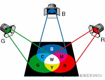
Image source: Baidu
2
Hyperspectral vs. multispectral imaging technology
Multispectral imaging has few spectral channels (a few to several dozen) and wide wavelength spacing, with no continuous spectral curve;hyperspectral imaging has stronger resolving power and can capture subtle spectral differences in materials, such as accurately distinguishing rock and mineral types in geological exploration.
Multispectral imaging is commonly used in remote sensing (land classification, vegetation surveys) and meteorological observation; its data volume is small and processing is easy. Hyperspectral imaging is suitable for fields with high requirements for compositional analysis, such as food additive detection and industrial quality inspection, but its data volume is large and processing is difficult.
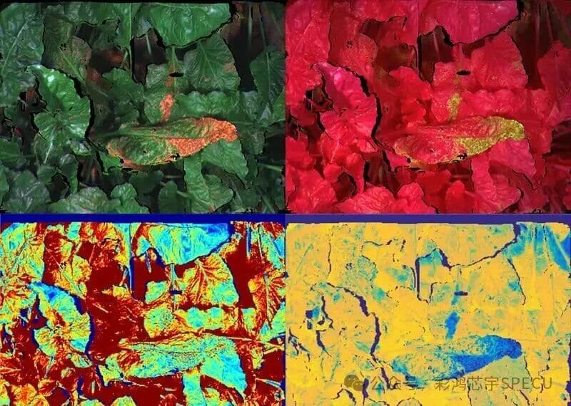
Image source: Sohu account
3
Hyperspectral vs. thermal imaging technology
Thermal imaging detects infrared radiation to display temperature distribution and is unrelated to spectroscopy;hyperspectral imaging does not directly measure temperature and focuses instead on compositional analysis.The two can be combined—for example, in fire rescue, thermal imaging locates trapped people and fire sources, while hyperspectral imaging analyzes hazardous gases.
Most thermal images are black-and-white or false-color and contain little detail; they are suitable for nighttime security, power equipment inspection, and body temperature screening.Hyperspectral imaging can provide color images and rich spectral information for subsequent analysis.
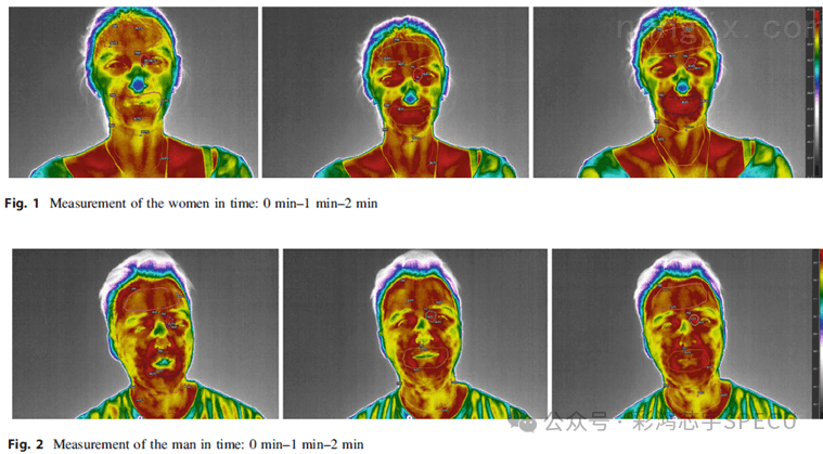
Image source: Agricultural Machinery Network
4
Hyperspectral vs. fluorescence imaging
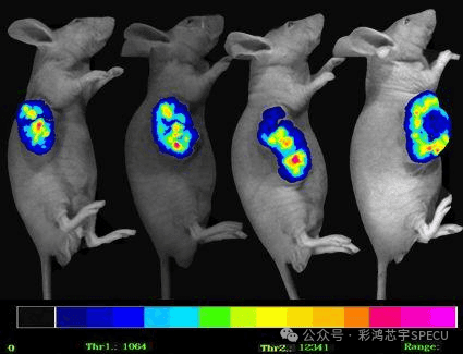
Fluorescence imaging requires excitation light to illuminate the target and captures the fluorescence signal it emits for imaging; at its core is “specific labeling” (for example, labeling cells with fluorescent probes);Hyperspectral imaging requires no labeling and identifies substances directly through spectral differences.
In terms of applications, fluorescence imaging is mainly used in biomedicine (cell tracking and tumor localization) and requires exogenous probes;hyperspectral imaging is suited to label-free scenarios, such as food mold detection and pigment analysis of cultural relics, and avoids probe interference.
In vivo mouse tumor study image
Image source: Instrument Information Network
5
Hyperspectral vs. terahertz imaging technology
Terahertz imaging uses terahertz waves (between infrared and microwaves) to penetrate non-metallic materials and detect internal structures; its core advantage is "non-destructive penetration." Hyperspectral imaging cannot penetrate; it focuses on surface and shallow-layer material composition.
Terahertz is mainly used for security screening (detecting concealed hazardous items) and semiconductor quality inspection (detecting internal defects); hyperspectral imaging is better suited to scenarios that require compositional analysis, such as qualitative analysis of pharmaceutical ingredients.
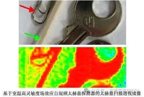
Image source: Baidu Baike
6
Hyperspectral vs. laser-induced breakdown spectroscopy imaging (LIBS)
LIBS uses a high-energy laser to strike the target and generate plasma, then identifies the material's composition by analyzing the plasma spectrum. Its defining feature is "micro-destructive testing," which requires damaging a tiny amount of the sample.Hyperspectral analysis is completely non-destructive.
LIBS is suitable for elemental analysis of geological ores and composition testing of metal materials; hyperspectral is better suited to scenarios with high sample integrity requirements, such as surface composition analysis of cultural relics and internal quality inspection of fruits and vegetables.
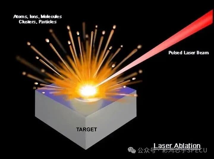
Image source: Baidu Baike
7
Hyperspectral vs. photoacoustic imaging technology
Photoacoustic imaging combines light absorption with ultrasonic detection, forming images from ultrasound signals generated when light irradiates a target. Its core capability is “deep imaging” (penetrating several centimeters of biological tissue); hyperspectral imaging has limited depth (mostly surface or thin layers).
Photoacoustic imaging is mainly used in biomedicine (such as early detection of breast tumors and cerebral vascular imaging);hyperspectral imaging is more accurate for analyzing the composition of superficial materials, such as spectral diagnosis of skin lesions.
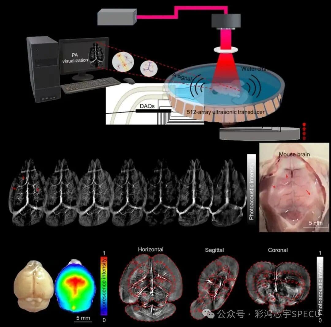
Image source: Sohu.com – New Optics
8
Hyperspectral vs. quantum dot imaging technology
Quantum dot imaging uses quantum dots as fluorescent probes, leveraging their high fluorescence intensity and narrow emission spectra for imaging; its core is “high-sensitivity labeling.” Hyperspectral imaging requires no labeling and relies on the intrinsic spectra of substances.
Quantum dot imaging is mainly used for biological cell imaging and LED displays; hyperspectral imaging suits scenarios where labeling is not possible, such as in situ detection of environmental pollutants.
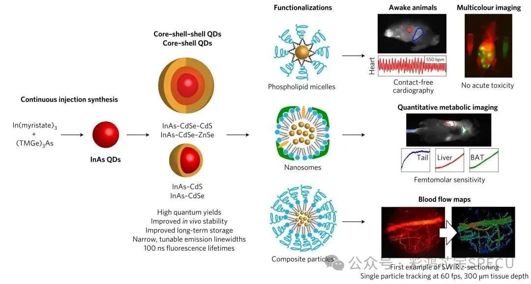
Image source: Sohu – Dongfang Shanguang
9
Hyperspectral vs. polarization imaging technology
Polarization imaging captures the polarization state of light (such as vibration direction and degree of polarization) to form images; its core is “distinguishing polarization differences in light” (such as identifying camouflaged targets and assessing object surface roughness);Hyperspectral imaging focuses on spectral differences.
Polarization imaging is mainly used in remote sensing (camouflaged target recognition) and medicine (retinal lesion detection in the fundus);hyperspectral imaging is better suited to identifying material composition,such as distinguishing different types of plastic.
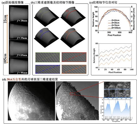
Image source: Institute of Computational Imaging
10
Hyperspectral vs. narrowband imaging technology
Narrow-band imaging selects a specific narrow wavelength range (usually 1–2 bands) for imaging; its core purpose is to “highlight specific targets” (such as suppressing background and enhancing lesion areas). Hyperspectral imaging covers a wide wavelength range with continuous bands.
Narrowband imaging is commonly used in medicine (polyp detection under gastrointestinal endoscopy) and security (low-light nighttime imaging);hyperspectral imaging can analyze multiple substances at once,such as detecting several additives in food simultaneously.
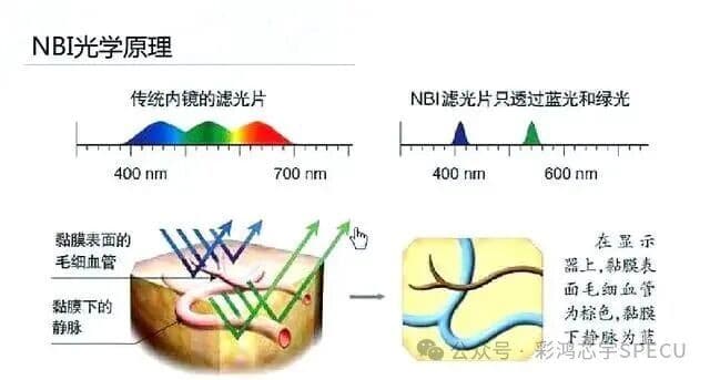
Image source: Baidu
11
Hyperspectral vs. holographic imaging technology
Holographic imaging records the amplitude and phase of light to reconstruct three-dimensional images, with 3D visualization as its core capability. Hyperspectral imaging captures only 2D images plus spectral information, with no 3D structure.
Holographic imaging is mainly used for artistic display and three-dimensional observation of biological samples; hyperspectral imaging focuses on compositional analysis, such as simultaneous detection of surface composition and defects on industrial products.
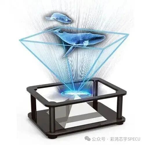
Image source: Baidu
12
Hyperspectral vs. X-ray imaging technology
X-ray imaging uses X-rays that pass through objects and forms images based on differences in absorption between tissues or materials. Its core principle is "penetration and density differentiation" (for example, inspecting internal structures or detecting metal foreign objects);hyperspectral imaging does not penetrate objects and emits no radiation.
X-ray imaging is often used in healthcare (fracture detection) and security screening (identifying foreign objects in luggage); hyperspectral imaging suits radiation-free scenarios that require compositional analysis, such as internal quality inspection of agricultural products.
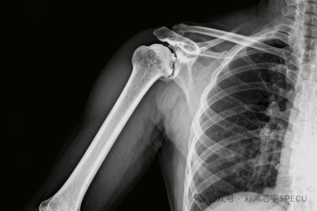
Image source: Doubao AI
Summary
RGB imaging is low-cost and easy to use, making it suitable for everyday scenarios;
Multispectral performs well in remote sensing and meteorology;
Thermal imaging expertise for temperature measurement;
Fluorescence and quantum dot imaging rely on labels and are primarily used in biomedicine.
Terahertz and X-ray are well suited to penetration inspection;
LIBS: minimally destructive elemental analysis;
Deep penetration for photoacoustic imaging
Polarization and narrowband imaging focus on specific optical properties;
Holographic imaging creates three-dimensional visualization.
Hyperspectral imaging, with its non-destructive measurement, continuous spectral bands, and precise compositional analysis as core strengths, is irreplaceable for precision detection in agriculture, food, healthcare, environment, industry, and other fields, and will integrate with other technologies in the future to expand into more application scenarios.